Privacy Policy for http://biologyhumanbody.blogspot.com/
If you require any more information or have any questions about our privacy policy, please feel free to contact us by email at mbokija@gmail.com.
At http://biologyhumanbody.blogspot.com/, the privacy of our visitors is of extreme importance to us. This privacy policy document outlines the types of personal information is received and collected by http://biologyhumanbody.blogspot.com/ and how it is used.
Log Files
Like many other Web sites, http://biologyhumanbody.blogspot.com/ makes use of log files. The information inside the log files includes internet protocol ( IP ) addresses, type of browser, Internet Service Provider ( ISP ), date/time stamp, referring/exit pages, and number of clicks to analyze trends, administer the site, track user’s movement around the site, and gather demographic information. IP addresses, and other such information are not linked to any information that is personally identifiable.
Cookies and Web Beacons
http://biologyhumanbody.blogspot.com/ does use cookies to store information about visitors preferences, record user-specific information on which pages the user access or visit, customize Web page content based on visitors browser type or other information that the visitor sends via their browser.
DoubleClick DART Cookie
.:: Google, as a third party vendor, uses cookies to serve ads on http://biologyhumanbody.blogspot.com/.
.:: Google's use of the DART cookie enables it to serve ads to users based on their visit to http://biologyhumanbody.blogspot.com/ and other sites on the Internet.
.:: Users may opt out of the use of the DART cookie by visiting the Google ad and content network privacy policy at the following URL - http://www.google.com/privacy_ads.html
Some of our advertising partners may use cookies and web beacons on our site. Our advertising partners include ....
Google Adsense
Commission Junction
Widget Bucks
Adbrite
Clickbank
Azoogle
Chitika
Linkshare
Amazon
Kontera
These third-party ad servers or ad networks use technology to the advertisements and links that appear on http://biologyhumanbody.blogspot.com/ send directly to your browsers. They automatically receive your IP address when this occurs. Other technologies ( such as cookies, JavaScript, or Web Beacons ) may also be used by the third-party ad networks to measure the effectiveness of their advertisements and / or to personalize the advertising content that you see.
http://biologyhumanbody.blogspot.com/ has no access to or control over these cookies that are used by third-party advertisers.
You should consult the respective privacy policies of these third-party ad servers for more detailed information on their practices as well as for instructions about how to opt-out of certain practices. http://biologyhumanbody.blogspot.com/'s privacy policy does not apply to, and we cannot control the activities of, such other advertisers or web sites.
If you wish to disable cookies, you may do so through your individual browser options. More detailed information about cookie management with specific web browsers can be found at the browsers' respective websites.
Thursday, January 6, 2011
Radioactive probes
Hemophiliacs suffer from defective Factor VIII, which can be detected in fetuses 20 weeks old. A more accurate test, which can also be administered earlier during pregnancy, involves the use of a radioactive probe (36 nucleotide RNA fragment) which hybridizes restriction fragments. The gene for hemophilia is 186,000 base pairs, and has 26 exons separated by 25 introns. Mutations in the gene can be detected by RFLPs. This technology has also been used to detect the single base-pair difference between normal and mutated beta-chains, a screen for sickle-cell anemia. A DNA probe has been developed that hybridizes with the gene for dystrophin. The previous screening for Duchenne Muscular Dystrophy was a sex screen, with option to abort a male. The new technique allows differentiation between the healthy and diseased male fetus, so parents have more information with which to make an informed choice (if they chose). The hybridization only occurs if the normal dystrophin gene is present, no hybridization occurs in the DMD sufferer.
Diagnosis of Human Genetic Diseases
Restriction enzymes, such as Hpa I were used in a study on sickle-cell anemia. The probe hybridized in normal hemoglobin with two fragments 7000 or 7600 nucleotides long. Sickle-cell hemoglobin had hybridization with a 13,000 nucleotide single sequence. A similar result has been obtained from amniocentesis studies, providing a tool to screen fetus and adult for sickle-cell. The markers where hybridization occurred are referred to as RFLPs (restriction-fragment-length polymorphisms). The longer fragment in sickle-cell individuals is interpreted as evidence of a mutation in the recognition sequence. Two nucleotide sequences close together on the same DNA molecule tend to stay together. In the sickle-cell DNA the beta-chain hemoglobin gene has become linked with another gene that somehow alters the recognition sequence at which Hpa I hybridizes. Heterozygotes will have both long and short fragments, while a single type (short or long) will occur in homozygous dominant and recessive, respectively.
Huntington's disease was studied by James F. Gusella and his research team, who used RFLPs to identify a marker. Testing a large library of human DNA fragments, Gusella et al. found the needle in the haystack. The enzyme used was Hind III. Four fragments have been identified in an American family that has members suffering from the disease. The presence of fragment A has been identified in individuals who suffer from (or will suffer from) Huntington's. Pattern A occurs in 60 percent of the population, as well as the Huntington's sufferers. A Venezuelan family of 3000 members is descended from a German sailor who had Huntington's. This family had a strong correlation between Fragment C and the disease. Pattern C is much less common among the general population in this country. Many individuals do not wish to know if they will develop this disease; Woody Guthrie's children have chosen not to be tested.
Cystic fibrosis (CF) has also been studied with RFLP technology. CF is the most common genetic disease in Caucasia
Sex-linked Traits
Color blindness afflicts 8% of males and 0.04 % of human females. Color perception depends on three genes, each producing chemicals sensitive to different parts of the visible light spectrum. Red and green detecting genes are on the X-chromosome, while the blue detection is on an autosome.
Hemophilia is a group of diseases in which blood does not clot normally. Factors in blood are involved in clotting. Hemophiliacs lacking the normal Factor VIII are said to have Hemophilia A, the most common form. Normal Factor VIII can be supplied at a high dollar and health risk cost, although the development of biotechnologically engineered Factor VIII produced by bacteria lessens the health risk. England's Queen Victoria was a carrier for this disease. The allele was passed to two of her daughters and one son. Since royal families in Europe commonly intermarried, the allele spread, and may have contributed to the downfall of the Russian monarchy (Czar Nicholas' son Alexei suffered from hemophilia A inherited from his mother who carried Victoria's genetic secret).


Muscular dystrophy is a term encompassing a variety of muscle wasting diseases. The most common type, Duchenne Muscular Dystrophy (DMD), affects cardiac and skeletal muscle, as well as some mental functions. DMD is an X-linked recessive occurring in 1 in 3500 newborns. Most sufferers die before their 20th birthday. In 1987, Louis Kunkel claimed to have isolated a protein, dystrophin, present in normal individuals (about 0.002 % of their muscle protein) but absent in two individuals with DMD. The lack of dystrophin is accompanied with a condition of muscle hardening known as fibrosis, which restricts blood supply to the muscle which then die. The gene technologies discussed in an earlier chapter have been employed to sequence and clone the dystrophin gene, which is the largest known human gene (some 2-3 million base pairs), with 60 exons and many large introns.
Hemophilia is a group of diseases in which blood does not clot normally. Factors in blood are involved in clotting. Hemophiliacs lacking the normal Factor VIII are said to have Hemophilia A, the most common form. Normal Factor VIII can be supplied at a high dollar and health risk cost, although the development of biotechnologically engineered Factor VIII produced by bacteria lessens the health risk. England's Queen Victoria was a carrier for this disease. The allele was passed to two of her daughters and one son. Since royal families in Europe commonly intermarried, the allele spread, and may have contributed to the downfall of the Russian monarchy (Czar Nicholas' son Alexei suffered from hemophilia A inherited from his mother who carried Victoria's genetic secret).
Muscular dystrophy is a term encompassing a variety of muscle wasting diseases. The most common type, Duchenne Muscular Dystrophy (DMD), affects cardiac and skeletal muscle, as well as some mental functions. DMD is an X-linked recessive occurring in 1 in 3500 newborns. Most sufferers die before their 20th birthday. In 1987, Louis Kunkel claimed to have isolated a protein, dystrophin, present in normal individuals (about 0.002 % of their muscle protein) but absent in two individuals with DMD. The lack of dystrophin is accompanied with a condition of muscle hardening known as fibrosis, which restricts blood supply to the muscle which then die. The gene technologies discussed in an earlier chapter have been employed to sequence and clone the dystrophin gene, which is the largest known human gene (some 2-3 million base pairs), with 60 exons and many large introns.
Human Allelic Disorders
The first Mendelian trait in humans was described in 1905 (brachydactly) by Dr. Farabee (no relation to your author). Now more than 3500 human genetic traits are known.
Albinism, the lack of pigmentation in skin, hair, and eyes, is also a Mendelian human trait. Homozygous recessive (aa) individuals make no pigments, and so have face, hair, and eyes that are white to yellow. For heterozygous parents with normal pigmentation (Aa), two different types of gametes may be produced: A or a. From such a cross 1/4 of the children could be albinos. The brown pigment melanin cannot be made by albinos. Several mutations may cause albinism: 1) the lack of one or another enzyme along the melanin-producing pathway; or 2) the inability of the enzyme to enter the pigment cells and convert the amino acid tyrosine into melanin.
Phenylketonuria (PKU) is recessively inherited disorder whose sufferers lack the ability to synthesize an enzyme to convert the amino acid phenylalanine into tyrosine Individuals homozygous recessive for this allele have a buildup of phenylalanine and abnormal breakdown products in the urine and blood. The breakdown products can be harmful to developing nervous systems and lead to mental retardation. 1 in 15,000 infants suffers from this problem. PKU homozygotes are now routinely tested for in most states. If you look closely at a product containing Nutra-sweet artificial sweetener, you will see a warning to PKU sufferers since phenylalanine is one of the amino acids in the sweetener. PKU sufferers are placed on a diet low in phenylalanine, enough for metabolic needs but not enough to cause the buildup of harmful intermediates.
Tay-Sachs Disease is an autosomal recessive resulting in degeneration of the nervous system. Symptoms manifest after birth. Children homozygous recessive for this allele rarely survive past five years of age. Sufferers lack the ability to make the enzyme N-acetyl-hexosaminidase, which breaks down the GM2 ganglioside lipid. This lipid accumulates in lysosomes in brain cells, eventually killing the brain cells. Although rare in the general population (1 in 300,000 births), it was (until recently) higher (1 in 3600 births) among Jews of eastern central European descent. One in 28 American Jews is thought to be a carrier, since 90% of the American Jewish population emigrated from those areas in Europe. Most Tay-Sachs babies born in the US are born to non-Jewish parents, who did not undergo testing programs that most US Jewish prospective parents had.
Sickle-cell anemia is an autosomal recessive we have discussed in other sections. Nine-percent of US blacks are heterozygous, while 0.2% are homozygous recessive. The recessive allele causes a single amino acid substitution in the beta chains of hemoglobin. When oxygen concentration is low, sickling of cells occurs. Heterozygotes make enough "good beta-chain hemoglobin" that they do not suffer as long as oxygen concentrations remain high, such as at sea-level.
Human chromosomal abnormalities
A common abnormality is caused by nondisjunction, the failure of replicated chromosomes to segregate during Anaphase II. A gamete lacking a chromosome cannot produce a viable embryo. Occasionally a gamete with n+1 chromosomes can produce a viable embryo.
In humans, nondisjunction is most often associated with the 21st chromosome, producing a disease known as Down's syndrome (also referred to as trisomy 21). Sufferers of Down's syndrome suffer mild to severe mental retardation, short stocky body type, large tongue leading to speech difficulties, and (in those who survive into middle-age), a propensity to develop Alzheimer's Disease. Ninety-five percent of Down's cases result from nondisjunction of chromosome 21. Occasional cases result from a translocation in the chromosomes of one parent. Remember that a translocation occurs when one chromosome (or a fragment) is transferred to a non-homologous chromosome. The incidence of Down's Syndrome increases with age of the mother, although 25% of the cases result from an extra chromosome from the father. Click here to view a drawing (from Bioweb) of a karyotype of Down's syndrome.
Sex-chromosome abnormalities may also be caused by nondisjunction of one or more sex chromosomes. Any combination (up to XXXXY) produces maleness. Males with more than one X are usually underdeveloped and sterile. XXX and XO women are known, although in most cases they are sterile. What meiotic difficulties might a person with Down's syndrome or extra sex-chromosomes face?

Kleinfelter's syndrome (click here to view a karyotype from Bioweb) How does this differ from the normal karyotype?
Turner's syndrome (click here to view a karyotype from Bioweb) How does this differ from the normal karyotype?
Chromosome deletions may also be associated with other syndromes such as Wilm's tumor.
Prenatal detection of chromosomal abnormalities is accomplished chiefly by amniocentesis. A thin needle is inserted into the amniotic fluid surrounding the fetus (a term applied to an unborn baby after the first trimester). Cells are withdrawn have been sloughed off by the fetus, yet they are still fetal cells and can be used to determine the state of the fetal chromosomes, such as Down's Syndrome and the sex of the baby after a karyotype has been made.
In humans, nondisjunction is most often associated with the 21st chromosome, producing a disease known as Down's syndrome (also referred to as trisomy 21). Sufferers of Down's syndrome suffer mild to severe mental retardation, short stocky body type, large tongue leading to speech difficulties, and (in those who survive into middle-age), a propensity to develop Alzheimer's Disease. Ninety-five percent of Down's cases result from nondisjunction of chromosome 21. Occasional cases result from a translocation in the chromosomes of one parent. Remember that a translocation occurs when one chromosome (or a fragment) is transferred to a non-homologous chromosome. The incidence of Down's Syndrome increases with age of the mother, although 25% of the cases result from an extra chromosome from the father. Click here to view a drawing (from Bioweb) of a karyotype of Down's syndrome.
Sex-chromosome abnormalities may also be caused by nondisjunction of one or more sex chromosomes. Any combination (up to XXXXY) produces maleness. Males with more than one X are usually underdeveloped and sterile. XXX and XO women are known, although in most cases they are sterile. What meiotic difficulties might a person with Down's syndrome or extra sex-chromosomes face?
Kleinfelter's syndrome (click here to view a karyotype from Bioweb) How does this differ from the normal karyotype?
Turner's syndrome (click here to view a karyotype from Bioweb) How does this differ from the normal karyotype?
Chromosome deletions may also be associated with other syndromes such as Wilm's tumor.
Prenatal detection of chromosomal abnormalities is accomplished chiefly by amniocentesis. A thin needle is inserted into the amniotic fluid surrounding the fetus (a term applied to an unborn baby after the first trimester). Cells are withdrawn have been sloughed off by the fetus, yet they are still fetal cells and can be used to determine the state of the fetal chromosomes, such as Down's Syndrome and the sex of the baby after a karyotype has been made.
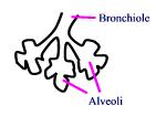
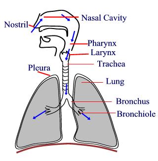
Human Respiratory system
1. Exchange of air occurs in _______ which are also known as 'air sacs'.
Ο Alveoli
Ο Alveolar ducts
Ο Bronchi
Ο Bronchioles
Answer
2. Identify bronchus in this diagram.
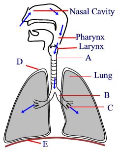
Ο A
Ο B
Ο C
Ο D
Answer
3. Write the correct sequence of the pathway through which air travels after entering the body.
A. Larynx, pharynx, trachea bronchioles
B. Pharynx, larynx, trachea, bronchioles
C. Pharynx, larynx, bronchioles, trachea
D. Pharynx, trachea, larynx, bronchioles
Ο A
Ο B
Ο C
Ο D
Answer
4. Which process does not occur in the nasal cavity?
A. Trapping of large foreign bodies
B. Exchange of gases
C. Humidification of inhaled air
D. Warming of inhaled air
Ο A
Ο B
Ο C
Ο D
Answer
5. Normal rate of respiration in an adult human being is _______ times/ minute.
A. 10-12
B. 12-14
C. 16-18
D. 22-24
Ο A
Ο B
Ο C
Ο D
Answer
6. Spirometer measures:
A. Capacity of lungs
B. Volume of air inhaled and exhaled
C. Residual air
D. All of these
Ο A
Ο B
Ο C
Ο D
Answer
7. Identify diaphragm in this diagram.
Ο B
Ο C
Ο D
Ο E
Answer
8. Complete the equation:
Glucose + Oxygen = _______ + Water + CO2
Ο Energy
Ο Sucrose
Ο Starch
Ο None of these
Answer
9. What is the leaf-like structure which prevents the entry of food into respiratory passages?
Ο Epiglottis
Ο Larynx
Ο Pharynx
Ο Tongue
Answer
10. Which part of the respiratory tract is also known as the voice box?
Ο Larynx
Ο Pharynx
Ο Trachea
Ο Epiglottis
Answer
11. Which cells of the blood carry oxygen to different parts of the body?
Answer
12. True or False: Trachea contains 16-20 'C'-shaped cartilaginous rings.
Ο True
Ο False
Answer
13. True or False: Breathing through the mouth is considered as good as breathing through the nose.
Ο True
Ο False
Answer
14. True or False: Exhalation and inhalation of air is known as "cellular respiration".
Ο True
Ο False
Answer
15. True or False: Two-layered membrane which covers the lungs is known as pericardium.
Ο True
Ο False
Ο Alveoli
Ο Alveolar ducts
Ο Bronchi
Ο Bronchioles
Answer
2. Identify bronchus in this diagram.
Ο A
Ο B
Ο C
Ο D
Answer
3. Write the correct sequence of the pathway through which air travels after entering the body.
A. Larynx, pharynx, trachea bronchioles
B. Pharynx, larynx, trachea, bronchioles
C. Pharynx, larynx, bronchioles, trachea
D. Pharynx, trachea, larynx, bronchioles
Ο A
Ο B
Ο C
Ο D
Answer
4. Which process does not occur in the nasal cavity?
A. Trapping of large foreign bodies
B. Exchange of gases
C. Humidification of inhaled air
D. Warming of inhaled air
Ο A
Ο B
Ο C
Ο D
Answer
5. Normal rate of respiration in an adult human being is _______ times/ minute.
A. 10-12
B. 12-14
C. 16-18
D. 22-24
Ο A
Ο B
Ο C
Ο D
Answer
6. Spirometer measures:
A. Capacity of lungs
B. Volume of air inhaled and exhaled
C. Residual air
D. All of these
Ο A
Ο B
Ο C
Ο D
Answer
7. Identify diaphragm in this diagram.
Ο B
Ο C
Ο D
Ο E
Answer
8. Complete the equation:
Glucose + Oxygen = _______ + Water + CO2
Ο Energy
Ο Sucrose
Ο Starch
Ο None of these
Answer
9. What is the leaf-like structure which prevents the entry of food into respiratory passages?
Ο Epiglottis
Ο Larynx
Ο Pharynx
Ο Tongue
Answer
10. Which part of the respiratory tract is also known as the voice box?
Ο Larynx
Ο Pharynx
Ο Trachea
Ο Epiglottis
Answer
11. Which cells of the blood carry oxygen to different parts of the body?
Answer
12. True or False: Trachea contains 16-20 'C'-shaped cartilaginous rings.
Ο True
Ο False
Answer
13. True or False: Breathing through the mouth is considered as good as breathing through the nose.
Ο True
Ο False
Answer
14. True or False: Exhalation and inhalation of air is known as "cellular respiration".
Ο True
Ο False
Answer
15. True or False: Two-layered membrane which covers the lungs is known as pericardium.
Ο True
Ο False
Human Digestive System

- Insoluble food has to be digested before it can be absorbed
- Chewing takes place in the mouth and breaks up the food
- Digestion starts digesting starch in the mouth. Amylase starts the process
- In the small intestine the starch has changed to glucose, so the digestion has stopped
- When starch is completely digested glucose is formed
- The digestion of protein starts in the stomach with an enzyme called pepsin
- The digestion of protein finishes in the small intestine
- The protein changes to amino acids after digestion
- In the small intestine, soluble food is absorbed into the blood
- In the rectum the water is taken out of the waste and the waste pushed together. This makes faeces.
Digestion: large nisoluble molecules are broken down into smaller soluble molecules
Absorption: small molecules are absorbed into the blood stream
Egestion: waste material passes out
Enzymes
- They are biological catalysts
- They speed up a reaction but remain unchanged at the end and can be used again
- Enzymes are proteins
- They are denatured (cease functioning) at high temperatures
- They are sensitive to pH
- Enzymes are apecific, only on enzyme will work with one substrate.
Carbohydrases
- Carbohydrases work on carbohydrates and break them down into simple sugars (e.g. Glucose). Amylase is an example of a carbohydrase
Proteases
- These work on proteins and break them down into amino acids. Examples include pepsin and trypsin.
Lipases
- These work on fats (lipids) and break them down into glycerol and fatty acids, for example lipase.
| Amylase | Pepsin | Trypsin | Lipase | |
|---|---|---|---|---|
| Where produced | Salivary glands and pancreas | Walls of stomach | Pancreas | Pancreas |
| Where functions | Mouth | Stomach | Small Intestine | Small Intestine |
| What they do | Break down of starch | Break down of protein | Break down of protein | Break down of fat |
What is Human Biology?
This subject allows students whose primary interest is in the human aspects of Biology to focus more closely on those areas than in the Biology A level. Because the Biology and Human Biology subject specifications have a considerable amount of common ground between them, students must elect to take one subject or the other; they may not take both. Please see Biology for further information.

Biological Symmetry
Animals exhibit one of two types of symmetry: bilateral, wherein an animal's right half is essentially a mirror image of its left half; or radial, wherein multiple lines of symmetry passing through a central point exist. (See figure below).
A snow board is a good example of a human-made object that exhibits bilateral symmetry (draw a line along the length of the ski to divide it in half and each side will be a mirror image of the other). A snow tube is a good example of a human-made object that exhibits radial symmetry (an infinite number of lines of symmetry can be drawn through the center of the tube as it lays flat on the snow).
To appreciate the functional difference between the two forms of symmetry, imagine yourself sliding down a hill on a snowboard and a tree appears in front of you. Now imagine the same scenario with a snowtube. Which object will be easier to steer away from the tree? Did you know that animals which move considerable distances under their own power are bilaterally symmetrical? Can you generate a hypothesis to explain this observation?
Additional examples of radial and bilateral symmetry in organisms are presented below. Can you identify which organisms exhibit which form of symmetry? How do each of these species move long distances in their environments?
 |
| Figure 1. A comparison of radial and bilateral symmetry in animals. |
 |  | |
| Figure 2. A snowboard (an example of a human-made bilaterally symmetrical object). | Figure 3. A snowtube (an example of a human-made radially symmetrical object). |
Additional examples of radial and bilateral symmetry in organisms are presented below. Can you identify which organisms exhibit which form of symmetry? How do each of these species move long distances in their environments?
 Shark |  Butterfly |  Jelly Fish |  |
Facial Asymmetry Experiments
| Table 1. Distance between the birdge of the nose and the left pupil (Lpupil), bridge of the nose and right pupil (Rpupil), bridge of the nose and left orbit (Lorbit), and bridge of the nose and right orbit (Rorbit). Key: Sex, 1=male and 2=female; Sightedenes, 1=normal, 2=near, 3=far, 4=astigmatism, and 5=other; Eye Dominance, 1=right, 2=left. | |||||||||
| Participant Number | Sex | Age | Sightedness | Handedness | Eye Dominance | Lpupil (mm) | Rpupil (mm) | Lorbit (mm) | Rorbit (mm) |
| 3129801 | 1 | 58 | 2 | 2 | 1 | 37 | 37 | 56 | 57 |
| 3129802 | 1 | 59 | 4 | 1 | 1 | 35 | 35 | 57 | 56 |
| 3129803 | 1 | 52 | 2 | 2 | 2 | 32 | 30 | 48 | 46 |
| 3129804 | 2 | 47 | 3 | 1 | 1 | 34 | 31 | 50 | 49 |
| 3129805 | 2 | 21 | 2 | 1 | 2 | 27 | 29 | 49 | 50 |
| 3129806 | 2 | 61 | 4 | 1 | 2 | 37 | 35 | 57 | 55 |
| 3129807 | 1 | 33 | 1 | 2 | 2 | 35 | 35 | 58 | 56 |
| 3129808 | 2 | 35 | 2 | 1 | 2 | 32 | 30 | 54 | 56 |
| 3129811 | 2 | 18 | 2 | 1 | 2 | 31 | 29 | 50 | 50 |
| 3129812 | 1 | 36 | 2 | 2 | 1 | 35 | 35 | 55 | 60 |
| 3129813 | 1 | 20 | 1 | 1 | 1 | 30 | 34 | 48 | 49 |
| 3129814 | 1 | 19 | 1 | 1 | 1 | 33 | 33 | 54 | 54 |
| 3129815 | 2 | 21 | 5 | 1 | 1 | 32 | 31 | 53 | 52 |
| 3129816 | 2 | 19 | 2 | 1 | 2 | 29 | 28 | 46 | 47 |
| 3129817 | 1 | 21 | 1 | 1 | 1 | 33.5 | 36.5 | 55 | 54 |
| 3129818 | 1 | 20 | 2 | 1 | 1 | 31 | 32 | 51 | 51 |
| 3129819 | 1 | 19 | 2 | 1 | 1 | 30.5 | 33.5 | 49.5 | 51.5 |
| 3129820 | 1 | 21 | 2 | 1 | 2 | 32 | 31 | 57 | 54 |
| 3129821 | 1 | 25 | 1 | 1 | 2 | 37 | 34.5 | 53 | 54 |
| 3129822 | 2 | 20 | 2 | 1 | 2 | 28 | 26 | 50 | 52 |
| 3129823 | 1 | 24 | 1 | 1 | 1 | 31 | 34 | 54 | 55 |
| 3129824 | 1 | 43 | 4 | 1 | 1 | 33.2 | 30.1 | 51.8 | 48.8 |
| 3129825 | 2 | 21 | 2 | 1 | 1 | 29.5 | 29 | 48 | 46 |
| 3129826 | 1 | 45 | 1 | 1 | 3 | 35 | 35 | 56 | 55 |
| 3129827 | 2 | 18 | 1 | 1 | 1 | 35 | 35 | 55 | 55 |
| 3129828 | 2 | 42 | 4 | 1 | 1 | 33 | 31 | 53 | 52 |
| 3129829 | 2 | 34 | 4 | 1 | 1 | 34 | 34 | 56 | 55 |
| 3129830 | 1 | 19 | 1 | 1 | 1 | 29 | 30 | 50 | 49 |
| 3129831 | 1 | 18 | 1 | 1 | 2 | 32 | 34.5 | 50 | 52.1 |
| 3129832 | 2 | 19 | 2 | 1 | 1 | 30.5 | 32 | 50.2 | 51 |
| 3129833 | 2 | 39 | 1 | 1 | 2 | 32 | 34 | 55.5 | 55 |
| 3129834 | 2 | 31 | 2 | 1 | 1 | 35 | 34 | 50 | 52 |
Human Body Symmetry

General biology textbooks list symmetry asa key concept used by scientists to differentiate various groups of animals. Authors of these texts usually state that all animals except sponges have their body parts arranged along an axis. This arrangement may be one of radial symmetry, wherein parts are arranged around a central axis, or bilateral symmetry, wherein an animal's right half is a mirror image of its left half. Humans are placed in the group of bilaterally symmetrical animals (Phylum Chordata). But, how bilaterally symmetrical are we? Is our left half a true mirror image of our right half?
A quick tour of one's internal anatomy reveals a lack of complete bilateral symmetry because we only have one heart and it is located in the left half of our thoracic cavity. Even the heart itself, with its four chambers, has a left ventricle that is substantially larger than the right ventricle.
What do you think about our external anatomy? Is our left side a mirror image of our right side? Can you design an experiment to answer this question? Can you think of other symmetry questions that could be addressed from an examination of external human anatomy?
Check some of the links below to learn how we have been investigating this problem and review some of the data we have collected. In fact, why don't you start measuring your body and let me [Buzz Hoagland

Human Biology

Human Biology
Learning the basics of human biology can be a complicated process. However, although difficult, biology genetics are considered to be a very interesting field to research, because of the nature of the work. Human biology is known as the scientific study of the body itself: how it works and what its made of. Unlike many other areas of biology which combines several areas into one, human biology concentrates specifically on people.
• The systems of the human body is considered to be part if its structure. Studying human biology is based on studying the bodily structure of a person. The building block of a living organism is called a cell. Cells combine to form larger structures called tissue. When learning about human biology, an individual may be required to do research with a microscope to look at these cells and tissues. A microscope is considered to be the most important tool to use when learning about human biology and biology genetics.
• One of the first things that an individual learns about human biology is that there are ten systems within the human body. These are the skeletal system, the excretory system, the reproductive system, the muscular system, the digestive system, the nervous system, the respiratory system, the immune system, and the endocrine system. A human being needs all those systems to be working in order to live, with the exception of the reproductive system. Human biology is often studied to learn about the causes, effects, and possible treatments of different diseases. Diseases will attack one of these systems when they begin to form. Some effects of a disease will be minor and others serious: this depends on the placement and the strength of the disease.
• Human biology concentrates on how the bodies’ cells, tissues, organs, and systems work together to form a human body. In biology, genetics is the type of study that covers the heredity and evolution of all life forms. Genetics is a cornerstone of human biology knowledge and study. Scientists have discovered that humans mostly all have the similar characteristics of things like eyes, a mouth and a nose. But in biology, genetics control how individuals look and live. Human biology relies particularly heavily on genetics since a humans genes determine peoples’ differences like eye color and hair color.
When studying biology, genetics and human biology is two complicated yet important subjects. Understanding how the different systems within the human body reacts with each other is an important part of understanding any basic knowledge of human biology.
Revealing the Target

HIV protease is a symmetrical molecule with two equal halves and an active site near its center. Molecular models of HIV protease in this chapter were generated by Alisa Zapp Machalek
With the structure of HIV protease at their fingertips, researchers were no longer working blindly. They could finally see their target enzyme—in exhilarating, color-coded detail. By feeding the structural information into a computer modeling program, they could spin a model of the enzyme around, zoom in on specific atoms, analyze its chemical properties, and even strip away or alter parts of it.

Knowing that HIV protease has two symmetrical halves, pharmaceutical researchers initially attempted to block the enzyme with symmetrical small molecules. They made these by chopping in half molecules of the natural substrate, then making a new molecule by fusing together two identical halves of the natural substrate.
These strategies worked in the case of HIV protease inhibitors. "I think it's a remarkable success story," says Dale Kempf, a chemist involved in the HIV protease inhibitor program at Abbott Laboratories. "From the identification of HIV protease as a drug target in 1988 to early 1996, it took less than 8 years to have three drugs on the market." Typically, it takes 10 to 15 years and more than $800 million to develop a drug from scratch.
 The structure of HIV protease revealed a crucial fact—like a butterfly, the enzyme is made up of two equal halves. For most such symmetrical molecules, both halves have a "business area," or active site, that carries out the enzyme's job. But HIV protease has only one such active site—in the center of the molecule where the two halves meet.
Pharmaceutical scientists knew they could take advantage of this feature. If they could plug this single active site with a small molecule, they could shut down the whole enzyme—and theoretically stop the virus' spread in the body.
Several pharmaceutical companies started out by using the enzyme's shape as a guide. "We designed drug candidate molecules that had the same two-fold symmetry as HIV protease," says Kempf. "Conceptually, we took some of the enzyme's natural substrate [the molecules it acts upon], chopped these molecules in half, rotated them 180 degrees, and glued two identical halves together."
To the researchers' delight, the first such molecule they synthesized fit perfectly into the active site of the enzyme. It was also an excellent inhibitor—it prevented HIV protease from functioning normally. But it wasn't water-soluble, meaning it couldn't be absorbed by the body and would never be effective as a drug.

A drug candidate molecule must pass many hurdles to earn the description "good medicine." It must have the best possible activity, solubility, bioavailability, half-life, and metabolic profile. Attempting to improve one of these factors often affects other factors. For example, if you structurally alter a lead compound to improve its activity, you may also decrease its solubility or shorten its half-life. The final result must always be the best possible compromise.
ANATOMY

The anatomy course fulfills a general education science requirement and serves as a gateway course to the nursing program. This course is a survey of the gross anatomy and histology of the major human organ systems including the skeletal and muscle systems, the digestive, circulatory, respiratory, excretory, nervous,
endocrine and reproductive systems. Laboratory work includes observation of models, and of human organs and tissues. A human cadaver is used for demonstration of musculature and digestive, respiratory, circulatory, and urogenital systems.
endocrine and reproductive systems. Laboratory work includes observation of models, and of human organs and tissues. A human cadaver is used for demonstration of musculature and digestive, respiratory, circulatory, and urogenital systems.
Culture in HAT media
Culture in HAT media

- Unfused myeloma cells (HGPRT-) die out because they cannot make purines.
- Unfused spleen cells (containing B-cells) die out because they have a limited life span
- The only cells that will survive are the fused B-cell: myeloma cells = hybridomas
2. Test fused cells for antibodies by ELIZA
3. Subculture and clone positives.
4. Test supernatants for antibodies
5. "Expand' or scale up the best clone (all derived from a single cell = monoclonal)
3. Subculture and clone positives.
4. Test supernatants for antibodies
5. "Expand' or scale up the best clone (all derived from a single cell = monoclonal)
6. Hybridoma cultures producing monoclonal antibodies can be maintained indefinitely in vitro or in vivo
7. MAbs can be developed into drugs, and injected into a person to seek out, bind, and target for destruction the antigen against which the antibody was raised.
The combination of antibody specificity (B cell) + unlimited proliferation (myeloma):
Because the human body eventually develops an immune response against the monoclonal antibody (made from a mouse B-cell), most monoclonal antibody-based drugs today are so-called humanized monoclonal antibodies: the mouse antibody is carefully digested to release just the antigen-binding variable region, which is then swapped into a human antibody missing its variable region. This prevents an immune response to the antibody itself.
A fully humanized monoclonal antibody has the antigen binding murine complementarity-determining regions interspersed within the variable regions of the light (light gray) and heavy (dark gray) chains of the Fab portion of the engineered antibody. A chimeric antibody has the entire antigen-binding murine component of the variable region of the Fab section is maintained integrally (handout).
See figures from Access Excellence and for humanized monoclonal antibodies.

The "Perfection" Of Biology
Creation Argument #5: The "Perfection" Of Biology
This argument goes something like "Nature is so complex, and works flawlessly with all its various parts, that it must have been created." At first glance, this would appear to have some merit of truth. Things do appear complex in nature and many things must work together in order to properly function. However, a closer inspection reveals that things are far from designed and even farther from being designed well. There are numerous instances that are explained well with evolution, and not explained at all by creation.
As a quick aside, I must talk about what it means to "explain" something in a theory. As I said previously, a theory is just a story that comprehensively accounts for all observable data. For data not yet observed, a theory must also be predictive, in that what the theory predicts for one situation must predict something similar for a similar situation, even if data has not been observed for it yet. For example, before humanoid fossils were discovered, back in the time when Darwin first proposed his theory, his theory still predicted their existence at some point in history. Whether we find the fossils or not is irrelevant, because most things do not fossilize. But it does predict something that we should be looking for, and indeed we have since found them. That is a powerful theory. The hypothesis of creation fails at this point, because it does not explain data, nor is it predictive at all. I'll explain some instances where nature is not optimal, where it is far from perfect, and where the prediction of a creator or designer fails.
Let's start with something very complex - the human eye. Many creationists will argue that the eye must be created, for what is the use of half an eye? With just briefly addressing this point to say that "half an eye" actually could and does retain and evolutionary survival advantage, what is important here is to realize that the photoreceptors of the eye are actually backwards. If it were designed, it would have been designed in the least optimal way possible. Evolutionarily, we can say that it is a "mistake" but one that conferred some advantage to the organism, and thereby retaining the genetic code for an eye (or proto-eye). Thus, the mistake was kept, and slowly improved upon, until the genetics were able to completely compensate for the original mistake.

As you can plainly see, the rods and cones are furthest away from the light as possible. The light actually must pass through the nerves (ganglion) to get to the receptors, which then are excited by the photons, and transmit their signal back through the nerves, down the optic nerve and into the brain. However, since the receptors are inverted, the optic nerve must travel through an area at the back of the eye, thereby creating our blind spot. This could all be avoided if the receptors were facing the "correct" way. In addition, after the optic nerve travels to the brain, it actually splits and crosses with the optic nerve of the other eye. This is surely do to the evolution of our brain to handle such information, but is a poor design nevertheless.
Here's another example: the brain produces a fluid called cerebrospinal fluid (CSF), which flows around the brain. It is very important for maintaining hydrostatic pressure and problems can cause hydroencephalitis, both through infection as well as congenitally. The only problem is, this fluid, so critical to the brain's function flows out through the cerebral aquaduct, which is a tiny little hole buried deep in the brain in between the two hemispheres. This spot could not be in a worse place, nor could the hole be any smaller to properly function. No rational designer would have put it there.
Example #3: The vague nerve (especially in giraffes). This nerve goes to the thyroid, but one of the nerves actually makes a huge detour all the way down and around the aorta, before returning back to its destination. In giraffes, this detour is huge! Why would any designer make this happen? They wouldn't. Of course. Evolution would explain it simply by saying that the evolutionary cost to extent the nerve was less than the cost of redesigning it completely.

Example #4: A protein in the human body called HIF-1. This protein is made, only to be immediately degraded in normal oxygenated conditions, and functions only in hypoxic conditions. No designer would built something to be torn down, just so that in the rare event of needing it, it would be there. Evolutionarily, there must be a tradeoff between the energy spent to have it versus the cost of not having it. In this sense, it is possible that local hypoxic conditions occur with enough frequency to retain constitutive expression of HIF-1.
I could go on and on, but the point has been made. Things function, but that is a far cry from saying they are designed. Evolution favors improved function, not necessary improved design. In other words, the design could actually get "worse", or farther from what would be ideal if designed from the ground up, but is better in the sense that it conveys some benefit in function from the previous version. We see this exact thing with the internet now. If we were to look at the internet today, we would say that it is far from what would be designed, if someone was designing it right now. But it was present in an original form in the '70s, and has subsequently been modified. Now, the internet is far too complex for anyone to go back and redesign it, because that would take far more energy than making constant small improvements. This is the same for evolution, because evolution works by a series of gradual modifications of an original form. The idea of creation does not favor any change at all, because a designer would have (should have) already supplied the organism with the most beneficial gene set possible. Anything less would require an imperfectly beneficent designer. As with all other areas, we see change, we see improvement, and both of these ideas are not explained by creation at all.
Health and disease
Investigate the science behind leading a healthy lifestyle and the major diseases of the body
 |
These resources are suitable for human biology classes and lessons that focus on nutrition, keeping fit and the connections between diet and disease.
Worksheets include structured practice of the language skills that students need to feel fully confident with the subject matter.
Worksheets include structured practice of the language skills that students need to feel fully confident with the subject matter.
Language support activities include:
* vocabulary and comprehension exercises
* speaking activities
* error correction work
* pronunciation practice
* vocabulary and comprehension exercises
* speaking activities
* error correction work
* pronunciation practice
Endocrine System Overview
The endocrine system, also referred to as the hormone system, is found in all mammals, birds, and fish. It is made up of:
- Glands located throughout the body.
- Hormones (i.e., chemical messengers) that are made by the glands and released into the bloodstream or the fluid surrounding cells.
- Receptors in various organs and tissues that recognize and respond to the hormones.
Hormones are released by glands and travel throughout the body searching for cells that contain matching receptors-proteins within the target cell or located on the surface of the target cell. The hormone binds with the receptor, much like a key would fit into a lock to unlock a door. The hormones, or keys, need to find compatible receptors, or locks, to work properly. Although hormones reach all parts of the body, only target cells with compatible receptors are equipped to respond. Once a receptor and a hormone have bonded, the receptor carries out the hormone's instructions by either altering the cell's existing proteins or turning on genes that will build a new protein. Both of these actions create reactions throughout the body. Researchers have identified more than 50 hormones in humans and other vertebrates.
The endocrine system regulates all biological processes from the conception of an organism through adulthood and into old age regulating many functions of a body, including metabolism, blood sugar levels, growth and function of the reproductive system, and the development of the brain and nervous system. The female ovaries, male testes, and pituitary, thyroid, and adrenal glands are all endocrine glands.
 From Biology: Principles and Explorations, Teaching Transparencies. Copyright 1996 by Holt, Rinehart and Winston. All rights reserved. Reprinted by permission of the publisher. |
The EPA's Endocrine Disruptor Screening Program focuses on the estrogen, androgen, and thyroid hormones. Estrogens, produced primarily by the ovaries and in small amounts by the adrenal glands, are the group of hormones responsible for female sexual development. Androgens are substances responsible for male sex characteristics. Testosterone, the sex hormone produced by the testicles, is an androgen. The thyroid gland secretes two main hormones, thyroxine and triiodothyronine, into the bloodstream that stimulate all the cells in the body and control many biological processes such as growth, reproduction, development, and metabolism.
Endocrine glands are located throughout the human body:
Hypothalamus - The hypothalamus links our endocrine and nervous systems together. The hypothalamus drives the endocrine system.
Pituitary gland - The pituitary gland receives signals from the hypothalamus. This gland has two lobes, the posterior and anterior lobes. The posterior lobe secretes hormones that are made by the hypothalamus. The anterior lobe produces its own hormones, several of which act on other endocrine glands.
Thyroid gland - The thyroid gland is critical to the healthy development and maturation of vertebrates and regulates metabolism.
Adrenal glands - The adrenal gland is made up of two glands: the cortex and medulla. These glands produce hormones in response to stress and regulate blood pressure, glucose metabolism, and the body's salt and water balance.
Pancreas - The pancreas is responsible for producing glucagon and insulin. Both hormones help regulate the concentration of glucose (sugar) in the blood.
Subscribe to:
Posts (Atom)